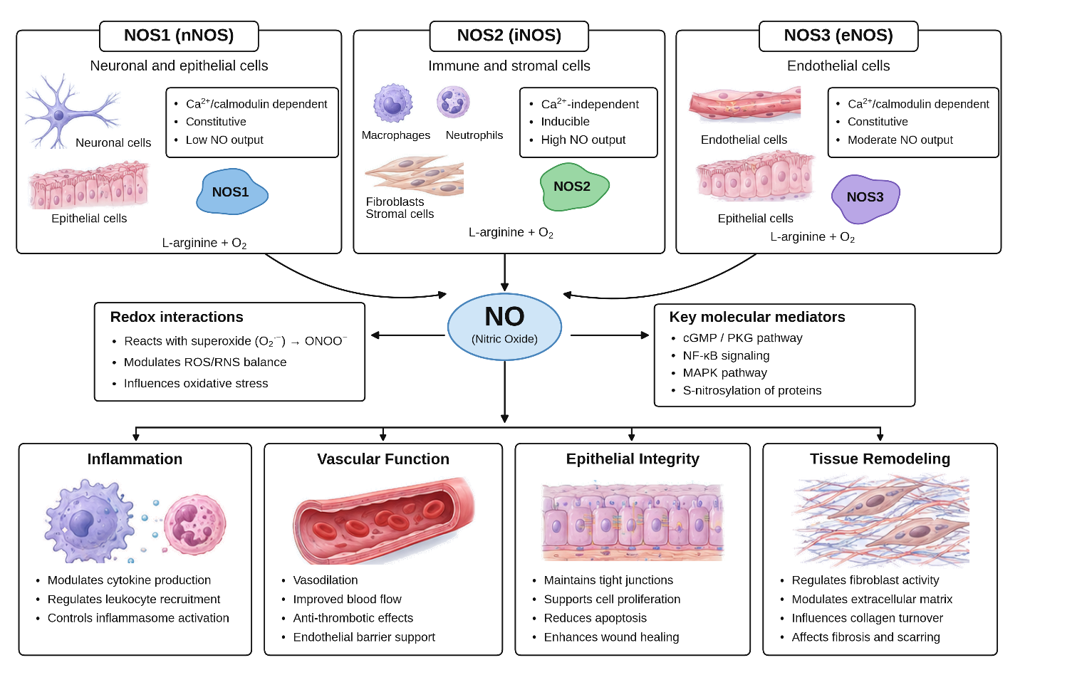
Figure 1: NOS isoforms and nitric oxide signaling in oral mucosal homeostasis and disease
Ejaz Ahmed1* | Ayisha Yousaf2 | Maryum Qasim3
1Medicine, IMPRS Berlin, Germany/ University of the Punjab/Shaikh Zayed Hospital, Lahore, Pakistan | 2Medical and Pharmaceutical Biotechnologies, University of Pavia, Italy | 3Department of Biochemistry, Kinnaird College for Women, Lahore, Pakistan
*Correspondence: Ejaz Ahmed (ejazahmedpatho@gmail.com)
Received: 29 September, 2026; Revised: 20 October, 2026; Accepted: 23 October, 2026; Published: 30 October, 2026
Nitric oxide (NO) signaling is involved in inflammatory processes, epithelial homeostasis, vascular function, and tissue remodeling, making it possible that expression of nitric oxide synthase (NOS) could be involved in oral mucosal and submucosal lesions. The aim of this narrative review is to discuss the reported expression patterns of NOS isoforms, their underlying molecular mechanisms, and the possible diagnostic and prognostic use. Oral leukoplakia and oral squamous cell carcinoma (OSCC) studies suggest that in certain cohorts there is increased expression of inducible NOS (NOS2/iNOS); however, these studies are limited by small sample size, methodological heterogeneity, and predominantly cross-sectional study design. There is still inconclusive evidence for oral submucous fibrosis and other potentially malignant disorders having consistent associations or independent prognosis. The distribution of NOS protein can be characterized by immunohistochemistry, which should be complemented by molecular and functional assays. Importantly, expression of NOS does not necessarily denote enzymatic activity or bioavailability of NO. The use of standardized methods and the assessment of isoforms, spatial analysis, and prospective multicenter validation are required before the establishment of NOS-derived biomarkers in routine oral pathology.
Keywords: Biomarkers; Carcinoma, Squamous Cell; Leukoplakia; Mouth Mucosa; Nitric Oxide.
Oral mucosal and submucosal lesions include reactive/inflammatory, oral cancer potentially malignant disorders (OPMDs), and oral squamous cell carcinoma (OSCC). he global burden is substantial; in 2022, an estimated 317,500 new cases of oral cavity cancer occurred worldwide 1. Although morphological characteristics are the most important feature, oral lesions with variable malignant potential, like leukoplakia and oral submucous fibrosis (OSMF), need to be evaluated based on clinical characteristics, histopathology, and molecular evidence 2
Nitric oxide (NO) is a transient molecule that plays a role in vascular regulation, immune function, and redox homeostasis. It is synthesized by neuronal, inducible, and endothelial NO synthase (NOS1, NOS2, and NOS3, respectively) 3. In particular, inflammation-associated NOS2/iNOS can produce a chronic production of NO; depending on concentration, duration, and surrounding oxidative environment, NO-related chemistry can affect cell signaling, DNA damage, angiogenesis, and tumor–stroma interactions. These mechanisms render the expression of NOS a possible molecular-pathology marker, but not a specific marker of dysplasia or malignant progression 4. Oral-tissue evidence is limited and is methodologically heterogeneous. It has been reported in a case-control immunohistochemical study that iNOS was stained in oral leukoplakia and OSCC, and that the expression of iNOS was correlated with some clinicopathological categories, but due to its cross-sectional nature, it cannot be used to predict the future 5. Therefore, it is important to differentiate between expression, enzymatic activity, availability of NO, and causal role in lesion progression.
The current narrative review examines NOS isoform expression in oral mucosal and submucosal lesions and summarizes possible associations with inflammatory, fibrotic, dysplastic, and OSCC lesions and appraises the tools for histopathologic and molecular assessment. It also explores the potential of NOS measurements to provide additional clinicopathological diagnostic and/or prognostic information that is reproducible. This narrative review was conducted to identify studies published between January 2020 and 13 October 2026 published data regarding the presence of NOS isoforms in normal oral mucosa, oral submucous fibrosis, oral potentially malignant disorders and oral squamous cell carcinoma. Controlled vocabulary and free-text terms were used to search PubMed/MEDLINE and citations, with the following terms being used: “nitric oxide synthase,” “iNOS,” “oral mucosa,” “oral submucous fibrosis,” “leukoplakia,” “dysplasia,” and “oral squamous cell carcinoma.” Primary tissue studies and relevant reviews were prioritized and studies compared according to the lesion, the isoform, the assay, and the clinicopathological associations. Results were synthesized narratively, taking into account conflicting results and limitations. This was not a systematic review.
NO Synthesis and Biological Functions: Nitric oxide (NO) is a short-lived signaling molecule involved in vascular regulation, cellular metabolism, and immune responses. Its biological effects depend on the local redox environment, the availability of reactive oxygen species, and the regulation of downstream signaling pathways 6. It is short-lived and can be diffused, so depends on location of production, local oxygenation, antioxidant capacity, and local reactive species for biological effects. In the oral mucosa, these pathways converge with the maintenance of the epithelial barrier, local perfusion, mucosal immunity, and tissue repair responses 7.
NOS Isoforms: Neuronal NOS (nNOS) and endothelial NOS (eNOS) are typically constitutively expressed and are subject to calcium–calmodulin regulation, though their levels of expression and regulation vary among different cell types. Whereas NO serves to regulate neuronal signaling in the nervous system, nNOS is important in non-neuronal tissues; eNOS is particularly important in endothelial cells to regulate vasodilation, microvascular function, and endothelial homeostasis. NOS2 (inducible NOS; iNOS), which is regulated mainly transcriptionally, can be induced by inflammatory cytokines and microbially triggered 8. Once expressed, the iNOS can produce NO more independently of transient calcium elevations 9. In oral tissues, the cellular source of NOS could be either the epithelial, vascular, immune or stromal compartment, and thus bulk-tissue measurements may not provide a reliable clue of the cells generating a signal 10. When interpreting the expression of lesions, it is therefore crucial to have specific location information.
Redox Context and Nitrosative Stress: NO doesn't always have protective or harmful effects. It can be involved in host defense and physiological signaling, but can also react with superoxide to generate peroxynitrite, a reactive oxidant that can modify proteins, lipids, and nucleic acids 11. The effects are concentration-dependent, time-dependent, and dependent on the balance between NO and superoxide. Under conditions of substrate deficiency or insufficient tetrahydrobiopterin, NOS may become uncoupled and produce superoxide rather than nitric oxide 12. These reactions provide a mechanistic rationale for investigating NOS in inflamed, fibrotic, dysplastic, and malignant oral tissues. However, increased NOS immunostaining does not necessarily indicate increased NO bioavailability or tissue injury. NOS expression, catalytic activity, and downstream redox products should therefore be considered related but distinct endpoints Figure 1 13.
Figure 1: NOS isoforms and nitric oxide signaling in oral mucosal homeostasis and disease
Normal Oral Mucosa and Inflammatory Lesions: Appropriate normal tissue controls are required for interpretation. A case–control study using immunohistochemistry found that the epithelial iNOS staining was not present in controls, but it was present in 73.3% of oral leukoplakia and 83.3% of oral squamous cell carcinoma (OSCC) samples 5. This indicates that there is a correlation between epithelial iNOS and disease, but not that NOS activity is always deficient in healthy mucosa. Distinct cellular sources can be missed by an individual overall score in inflamed lesions where the different compartments (epithelial, immune and stromal) contribute differently 14.
Oral Submucous Fibrosis: Oral submucous fibrosis (OSMF) is a progressive condition characterized by subepithelial fibrosis and epithelial changes and has been associated with a known, but variable, risk of malignant progression 15. However, the evidence for OSMF based upon tissues has been limited in recent years. Therefore, proposed relationships of iNOS-associated inflammation, vascular responses and epithelial–stromal remodeling should be considered as hypotheses rather than causal pathways. Studies should include the localization of NOS2 to epithelium, fibroblasts, endothelial cells and inflammatory infiltrates, and correlate expression with fibrosis grade, dysplasia and exposure history.
Oral Leukoplakia and Epithelial Dysplasia: Oral leukoplakia is the only confirmed potentially malignant lesion, while the evidence for the other lesions is limited. In the 2020 study, epithelial iNOS was found in 73.3% of leukoplakia samples, with this expression being associated with higher clinical stage and dysplasia grade of the disease 5. This is consistent with the correlation with lesion severity but not prediction of transformation, as the cross-sectional design did not yield a prospective risk discrimination. Another systematic review published in 2025 states that leukoplakia is not homogeneous, that non-homogeneous appearance and lesion size and location on the tongue and epithelial dysplasia affect the risk of transformation 16. NOS expression therefore needs to be compared with established clinicopathological variables to see if it provides additional information.
Verrucous Hyperplasia and Verrucous Carcinoma: The verrucous lesions, due to their architectural and behavioral characteristics, should be evaluated because they may not have all the characteristics of leukoplakia and OSCC. Post-2020 literature retrieved for review, however, failed to identify any strong human tissue studies that have been performed to determine isoform-specific NOS patterns in verrucous hyperplasia and verrucous carcinoma 17. The proposed changes in the localization or intensity of iNOS, therefore, should be viewed as an evidence gap and not as a result of older studies or extrapolated from traditional OSCC. The diagnosis of focal dysplasia or conventional invasive components requires consistent criteria and enough sampling of the comparative cohorts.
Oral Squamous Cell Carcinoma: Experimental and clinical investigations of the oral squamous cell carcinoma microenvironment have highlighted interactions among tumour cells, immune populations, and inflammatory signaling pathways that may contribute to metastatic progression 18. The work implicated iNOS+myeloid cells and CXCL5–CXCR2 signaling, but preclinical intervention does not validate iNOS staining as a clinical prognostic test. The 2020 tissue study also found that 83.3% of OSCC tissue samples were iNOS-positive, and there was a correlation between iNOS positivity and clinicopathological stage and differentiation 19. These results warrant continued investigation of epithelial and immune-cell NOS2, and its longitudinal and independent validation.
Comparative Synthesis: There is evidence indicating that iNOS is a potential correlate of oral epithelial pathology and not a validated marker of progression. Data were mostly focused on leukoplakia and OSCC, with insufficient data available for isoform-resolved data in OSMF and verrucous lesions. The evidence available for the disease lesions is summarized in Table 1 and the main conclusions and limitations of recent studies are stated.
| Study (Reference) | Lesion or research focus | Principal finding or contribution | Evidence classification |
|---|---|---|---|
| Singh et al., 2020 5 | Oral leukoplakia and OSCC | iNOS positivity was reported in 73.3% of leukoplakia and 83.3% of OSCC specimens in a participant case–control study. | Direct human oral-tissue evidence |
| Piacenza et al., 202211 | NO, superoxide and peroxynitrite | Examined redox-dependent mechanisms that can influence the biological consequences of NO production. | Biochemical and mechanistic evidence |
| Bouaoud et al., 2021 20 | Oral potentially malignant disorders and the immune microenvironment | Combined epithelial–stromal RNA sequencing and immunohistochemistry, including iNOS-positive M1 macrophages, to investigate immune changes during oral carcinogenesis. Macrophage gene-expression signatures were subsequently assessed in 86 human oral leukoplakia cases for associations with oral cancer-free survival. | Preclinical oral-tissue evidence with supporting human transcriptomic outcome data; iNOS staining was not directly validated in the human cohort. |
| Kavitha et al., 202221 | Oral submucous fibrosis | Reviewed immunohistochemical biomarkers investigated in OSMF and highlighted the heterogeneous evidence base. | Supporting review; not an independent NOS-expression cohort |
| Lundberg and Weitzberg, 202222 | NO signaling and disease biology | Reviewed physiological and pathological NO signaling, providing a framework for interpreting NOS-associated findings. | General mechanistic evidence |
| Goldsmith et al., 2024 23 | Immunohistochemical assay validation | Provided updated guidance on analytical validation of immunohistochemical assays relevant to the design of NOS biomarker studies. | Methodological evidence |
An important difference is that evidence of the same mechanism through broader cancer models does not guarantee the same mechanism in human oral mucosal disease.
Inflammation and Transcriptional Regulation: Cytokines can induce NOS2 expression through NF-κB and other cytokine-responsive transcriptional pathways. In chronically inflamed oral tissues, interactions between epithelial cells, macrophages, and other immune cells may contribute to sustained inflammatory signaling; however, these mechanisms require direct evaluation in oral lesions 24. The final product, NO, may affect the immune responses and cellular signaling and may have a different effect depending on the immune cells producing NO and the microenvironment. Immunoreactivity for NOS2 is, however, not sufficient for NF-κB activation or to specify the upstream stimulus. Because the pathway is demonstrated by the combination of staining intensity with the complementary evaluation of NOS2, inflammatory mediators, and related transcriptional activity, inferring pathway activation from staining intensity alone is not sufficient to demonstrate this pathway in oral lesions 25.
Oxidative and Nitrosative DNA Damage: NO reacts with reactive oxygen species to produce reactive nitrogen species, such as peroxynitrite, that can alter bases in DNA, proteins, and membrane lipids. These reactions can lead to genomic instability if the damage is more serious than the capacity of the cell to repair. Oxidative and nitrosative stress may also disrupt DNA-repair pathways and redox-sensitive signaling, which may affect the survival of genetically aberrant epithelial cells 26. However, the amount of NOS does not confirm the occurrence of DNA damage 27. Establishing this relationship requires direct measurements of relevant damage products, appropriate oxidative-stress controls and assessment of DNA-repair responses in the same tissue specimens.
Epithelial Proliferation, Apoptosis and Survival: NO can regulate cell-cycle, mitochondrial function, apoptosis and survival via concentration-dependent signaling. Exposure to lower or transient NO concentrations might induce a physiological signaling, while a continuous production of NO in an oxidizing environment might lead to cellular injury or change the survival of the tumor cells. Because of these opposing properties, it is not suitable to label iNOS as a blanket term of tumor promoters or tumor suppressors. The biological relevance of its presence in oral epithelial lesions should be considered in conjunction with dysplasia, its cellular localization, and independent evaluations of proliferation and apoptosis. These endpoints might also help to establish biological relevance but not, in the absence of longitudinal or experimental evidence, a causal role in malignant transformation 28.
Angiogenesis and Stromal Interactions: NO is involved in endothelial cell signaling and vascular remodeling, and tumor-associated inflammatory cells have the potential to interact with the stromal environment via cytokines and chemokines. In models of squamous cell carcinoma, iNOS activation of myeloid cells is associated with metastatic behaviour, warranting investigation of the interactions between inflammatory and tumor compartments 29. For oral lesions, however, the vascular density, localization of NOS isoforms and stromal signaling should be assessed individually prior to interpretation of the association as evidence of NOS-mediated angiogenesis. Spatially resolved measurements might be useful to assess what these processes are and whether NOS expression in the epithelium or stroma would be better 30.
Fibrosis and Extracellular Matrix Remodeling: Oral submucous fibrosis (OSMF) is the result of chronic inflammation and increased levels of extracellular matrix, leading to progressive fibrosis and changes in the architecture of the mucosa. The potential relationships among NO-related signaling and fibroblast activity, release of inflammatory mediators, and degradation of matrix are not well understood, however, in human OSMF tissue 31. The extrapolations of other fibrotic diseases cannot be made to oral submucosal fibrosis. It is also important to investigate NOS isoforms in conjunction with collagen deposition, markers of myofibroblasts, and matrix remodeling enzymes to assess if findings of association are functional or merely associated with established fibrotic changes Figure 2 32.
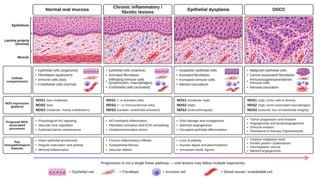
Figure 2: Conceptual histopathological framework for NOS expression across oral lesion progression
Immunohistochemistry: Immunohistochemistry (IHC) is especially useful to examine NOS expression in the context of maintaining the tissue architecture and allows for an examination of the localization of NOS isoforms in the epithelial, stromal, vascular, and inflammatory compartments. Validated antibodies, positive and negative controls, antigen retrieval, and staining protocols do account for reliable interpretation 33. The biological functions of NOS1, NOS2, and NOS3 are significantly different, and antibody specificity is particularly relevant when discriminating their functions. Staining intensity, percentage of positive cells, subcellular localization and immunoreactivity in different compartments of the histological sections should be documented. There may be some interpretive variability that is reduced by a prespecified scoring system, blinded assessment, and interobserver-agreement analysis. It is important to note that the positive IHC result is indicative of protein immunoreactivity, but not necessarily of enzyme activity or increased NO production 34.
Molecular and Functional Assays: Complementary techniques can enhance the information provided by tissue diagnosis. However, reverse-transcription quantitative polymerase chain reaction (RT-qPCR) only quantifies the amount of NOS transcript and does not provide information on protein localization or catalytic activity. In situ hybridization gives spatial data about transcripts, and enzyme-activity assays measure catalytic activity under specific experimental conditions 35. If available, measurements of nitrate, nitrite or other NO-related products can also give biochemical clues, but these can be affected by many biological and environmental factors and do not necessarily show which NOS isoform is responsible for the production of NO. Where possible, then, it is better to use spatial protein determinations in conjunction with transcript analysis and some functional measurements 36. However, a consensus among the independent methods supports the biological interpretation, while differences could be due to posttranscriptional regulation, heterogeneity of the tissue or discrepancies between expression level and enzyme activity.
Study Design and Statistical Interpretation: Interpretation is also subject to variables such as selection of cohorts, histopathological classification, and control of confounding variables. Studies should establish the inclusion criteria, differentiate a nondysplastic lesion from a graded epithelial dysplasia, and record exposures to tobacco and alcohol (if applicable). One of the essential things is to justify the samples size and have prespecified endpoints as well as adjust for any confounders to prevent unstable associations. There could be a risk of false positives for multiple comparisons between different NOS isoforms, staining compartments, and clinical subgroups. Where statistical analysis indicates that there are no grounds for prematurely converting a continuous or ordinal staining score into a positive/negative classification, this should be done. Proposed cutoffs should be tested in independent validation cohorts, and the results should be reproducible before they are used for clinical decision-making. Analytical validation of immunohistochemical assays is essential before their clinical application, particularly when staining results are intended to inform diagnostic or prognostic decisions 37. Table 2 provides an overview of the different ways in which NOS expression and activity can be assessed, and a discussion of the strengths and weaknesses of each approach.
| Study (Reference) | Assessment method or research focus | Principal contribution | Strengths and limitations |
|---|---|---|---|
| Goldsmith et al., 202423 | Immunohistochemistry (IHC) assay validation | Provides updated principles for validating antibody-based assays before clinical application. | Supports analytical validation; does not specifically validate NOS antibodies in oral lesions. |
| Webster et al., 202138 | IHC antibody specificity | Describes a weight-of-evidence approach to assessing antibody specificity in investigative studies. | Highlights the importance of orthogonal validation and appropriate controls; application is not NOS-specific. |
| Lundberg and Weitzberg, 2022 22 | NO signaling and biochemical interpretation | Explains the biological distinction between NO signaling and downstream physiological or pathological effects. | Provides mechanistic context; cannot substitute for direct measurement of NOS activity in oral tissue. |
| Piacenza et al., 2022 11 | Reactive nitrogen species and redox biology | Explains interactions between NO, superoxide and peroxynitrite. | Supports interpretation of oxidative and nitrosative endpoints; these products do not uniquely identify the responsible NOS isoform. |
| Singh et al., 2020 | IHC assessment of iNOS in oral leukoplakia and OSCC | Demonstrates the application of tissue-based iNOS immunostaining in human oral lesions. | Preserves tissue localization; cross-sectional findings cannot establish enzymatic activity or prospective prognostic value. |
| Silva Servato et al., 201939 | iNOS and nitrotyrosine assessment in normal oral mucosa, oral leukoplakia, and oral squamous cell carcinoma (OSCC) | Evaluated NOS2 mRNA using qPCR and assessed iNOS and nitrotyrosine using immunohistochemistry. Expression patterns were associated with oral carcinogenesis and several clinicopathological parameters. | Combines molecular and tissue-based evidence across different oral lesion categories; however, independent validation is required before these markers can be recommended for routine prognostic assessment. |
Diagnostic Relevance: NOS immunoreactivity could serve as additional data to the conventional histopathology in the evaluation of inflammatory and molecular features of oral mucosal lesions, especially when used in conjunction. Its diagnostic contribution is still under investigation, however. Potentially malignant disorders of the oral cavity are a diverse group of disorders, and expression of NOS may be a manifestation of inflammation, vascular responses, or changes to the epithelial microenvironment, and not necessarily dysplasia 40. Therefore, NOS staining should not replace morphological evaluation or established diagnostic criteria. Its potential role is to identify biological differences not fully captured by routine histopathological examination, provided that isoform-specific localization demonstrates reproducible and clinically relevant patterns 41.
Prognostic Relevance: To determine the prognostic value, evidence over the course of time is needed that baseline NOS measurements are linked to malignant transformation, recurrence, or disease progression. The difference in cross-section between leukoplakia and OSCC is not sufficient to establish the predictive performance, as they do not show that the change measured is pre-transformational. Future studies need to evaluate if the expression of NOS provides additional prognostic information over dysplasia grade, features of the lesion and exposure history. The reproducibility of a prognostic biomarker is a prerequisite for it to be a prespecified outcome or a biomarker that is independently validated 42.
Clinical Translation: A standardized and analytically validated assay and a reproducible threshold for scoring would be required for translation to clinical practice. While the recommendations given in the guidelines do not include validation of NOS testing in oral lesions, the update to the guideline for the 2024 edition of the College of American Pathologists (CAP) reiterates the necessity for assay-specific validation prior to clinical use 23. Future multicenter diagnostic accuracy, calibration, reproducibility, and incremental predictive value studies are needed with respect to conventional clinicopathological variables. The clinical utility can only be demonstrated by demonstrating the improvement of decision-making or patient outcomes through the use of NOS-informed risk assessment. Until the above are achieved, NOS expression should be used as a research biomarker and not as a sole diagnostic, surveillance, or treatment selection criterion.
The evidence currently available for NOS expression in oral mucosal and submucosal lesions is limited, and thus interpretation for clinical use is limited. First, there are limited human tissue studies available, and many of these studies are cross-sectional and cannot determine if alterations in NOS expression occur before or after dysplasia or before or after already existing pathological changes. Second, the specificity of antibody employed, staining techniques, scoring criteria and tissue processing all make it difficult to compare results from different studies. Even less biological specificity exists due to inconsistent separation of NOS isoforms and cellular compartments.
An important question that remains is how NOS expression relates to the functional NO signaling. The abundance of protein, the levels of transcripts, the catalytic activity and downstream levels of reactive N species are not equivalent biological endpoints and can lead to misleading conclusions if treated as such. Additionally, there is a problem of lesion heterogeneity, especially if inflammatory activity, fibrosis, epithelial dysplasia, and invasive carcinoma are found in the same specimen.
In third, lack of uniformity in the NOS-specific evidence, especially in terms of the longitudinal data available for oral submucous fibrosis and verrucous lesions. Oral leukoplakia is a highly heterogeneous condition, as evidenced by the wide range in malignant-transformation estimates 43. The improvement in risk prediction with NOS measurements over the known clinicopathological risk factors is unclear. This question should be considered in planning future investigations, but not as an assumption that a statistically significant association necessarily translates to clinical utility.
Future studies should progress from descriptive NOS immunostaining to integration in the characterization of oral lesions by NOS isoform. Multiplex immunohistochemistry and spatial transcriptomics might help to differentiate the NOS expression of dysplastic epithelium, fibroblasts, endothelial cells, and immune populations without altering the tissue architecture 44. Single-cell transcriptomic techniques could also help identify the cell sources and the corresponding signaling pathways, but the detection of the transcript does not imply the activity of the enzyme 45. These strategies are gaining significance in the field of oral cancer research, where spatial analysis is unravelling the interactions between the epithelial and stromal components and the complexities of cellular heterogeneity.
Prospective, multicenter cohorts should use harmonized assays and prespecified scoring criteria as well as documented exposure history to assess NOS1, NOS2 and NOS3. Protein localization should be coupled with the abundance of transcript, functional measurements and clinicopathological outcomes, if possible. Predictive models need to be validated independently and show incremental value over known risk factors before they are used in the clinic.
The expression of nitric oxide synthase is a plausible method of investigation of inflammatory, fibrotic, dysplastic and malignant changes in oral tissues. Current evidence favors the further investigation of inducible NOS (NOS2/iNOS) in oral leukoplakia and oral squamous cell carcinoma, but lacks the power to prove its stand-alone role. Interpretation should account for differences between protein expression and enzymatic activity and downstream NO effect. To assess whether NOS measurements add value to established clinicopathological variables for risk assessment there is a need for isoform-specific localization and standardized assays and prospective validation. NOS expression should continue to be considered an investigational biomarker until such evidence is obtained.
This work is published by Multidisciplinary Scholarly Advancement and Research MSAR Institute. © 2026 The Author(s). The full terms of Journal Publishing policies are available at https://msarjournals.com/index.php/index/open-access and incorporate the Creative Commons Attribution – Non Commercial (CC BY-NC 4.0) License https://creativecommons.org/licenses/by-nc/4.0/. By accessing the work you hereby accept the Terms. Non-commercial uses of the work are permitted without any further permission, provided the work is properly attributed. Publisher’s Note: MSAR Institute remains neutral with regard to jurisdictional claims in published maps and institutional affiliations, and assumes no liability for the scientific accuracy or clinical efficacy of the content herein, as they rest entirely with the authors.