Beyond Calcification: Emerging Pathological, Molecular, and Clinical Significance of Pineal Gland Lesions
DOI:
https://doi.org/10.68041/medsc.v1i1/02Keywords:
Brain Neoplasms, Calcinosis, Cysts, Melatonin, Neuroimaging, Pineal Gland, PinealomaAbstract
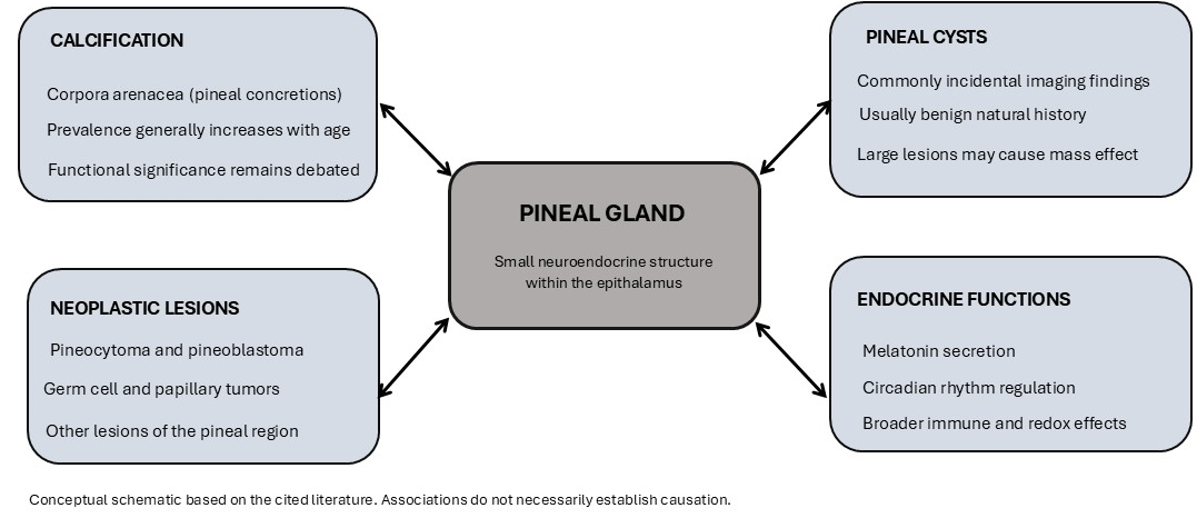
The pineal gland is a small neuroendocrine gland located in the middle of the epithalamus and was long thought to be a vestigial structure whose main clinical significance was in the diagnosis of calcification on imaging. The more streamlined perspective is quickly becoming unsustainable. In modern imaging, molecular pathology, and endocrine studies, the pineal region is now known to be the site of complex interrelationships between calcification, cyst formation, and the development of neoplasms, with melatonin physiology, circadian rhythms, and oncogenic signaling. The aim of this communication is to present the current knowledge regarding the pathological and molecular diversity of pineal lesions, and to discuss the longstanding controversy of the functional consequences of pineal calcification and the potential clinical consequences of imaging-pathological discordance. It is believed that this pineal gland requires resurgence of investigation, not simply because of the occasional radiographical finding, but for its possible role as a target for alterations in neurological and systemic disease.
References
1. Bukreeva I, Junemann O, Cedola A, Palermo F, Maugeri L, Provinciali GB, et al. Investigation of the human pineal gland 3D organization by X-ray phase contrast tomography. J Struct Biol. 2020;212(3):107659. https://doi.org/10.1016/j.jsb.2020.107659
2. Ikram J, Akram S. Exploring the Role of Oxidative Stress-Related Molecular Biomarkers in Cardiovascular Disease Progression and Therapeutic Modulations: A Systematic Analysis. J Biomol Pathog Ther. 2026;2(1):35-42. https://doi.org/10.68041/jbpt.v2i1/04
3. Zheng J, Song W, Zhou Y, Li X, Wang M, Zhang C. Cross-species single-cell landscape of vertebrate pineal gland. J Pineal Res. 2024;76(1):e12927. https://doi.org/10.1111/jpi.12927
4. Jalali N, Firouzabadi MD, Mirshekar A, Khalili P, Ravangard AR, Ahmadi J, et al. Cross-sectional analysis of potential risk factors of the pineal gland calcification. BMC Endocr Disord. 2023;23(1):49. https://doi.org/10.1186/s12902-023-01301-w
5. Junemann O, Bukreeva I, Otlyga DA, Cedola A, Fratini M, Saveliev SV. Human pineal gland involutionary process: new findings. J Gerontol A Biol Sci Med Sci. 2023;78(9):1558-60. https://doi.org/10.1093/gerona/glad091
6. Mutalik S, Tadinada A. Prevalence of pineal gland calcification as an incidental finding in patients referred for implant dental therapy. Imaging Sci Dent. 2017;47(3):175-80. https://doi.org/10.5624/isd.2017.47.3.175
7. Del Brutto OH, Mera RM, Arias EE, Rumbea DA, Patel V, Castillo PR. Automated measurement of pineal gland calcification volumes and sleep quality in adults living in costal Ecuador. Arq Neuropsiquiatr. 2026;84(01):001-5. https://doi.org/10.1055/s-0045-1814399
8. Al Mutawa M, Lange I, Kirsch M, Flöel A, Fleck S, Schroeder HW. Symptomatic pineal cysts without ventricular enlargement: diagnosis and treatment. Dtsch Arztebl Int. 2025;122(11):298. https://doi.org/10.3238/arztebl.m2025.0053
9. Masina R, Ansaripour A, Beneš V, Berhouma M, Choque-Velasquez J, Eide PK, et al. Surgical treatment of symptomatic pineal cysts without hydrocephalus—meta-analysis of the published literature. Acta Neurochir (Wien). 2022;164(1):61-77. https://doi.org/10.1007/s00701-021-05054-0
10. Fakhran S, Escott EJ. Pineocytoma mimicking a pineal cyst on imaging: true diagnostic dilemma or a case of incomplete imaging? AJNR Am J Neuroradiol. 2008;29(1):159-63. https://doi.org/10.3174/ajnr.A0750
11. Yuan S, Lin Y, Zhao Y, Du M, Dong S, Chen Y, et al. Pineal cysts may promote pubertal development in girls with central precocious puberty: a single-center study from China. Front Endocrinol (Lausanne). 2024;15:1323947. https://doi.org/10.3389/fendo.2024.1323947
12. Jiang Z, Allkanjari MS, Chung PE, Tran H, Ghanbari-Azarnier R, Wang DY, et al. Recent advances in pineoblastoma research: molecular classification, modelling and targetable vulnerabilities. Cancers (Basel). 2025;17(5):720. https://doi.org/10.3390/cancers17050720
13. Wu Z, Dazelle K, Abdullaev Z, Chung HJ, Dahiya S, Wood M, et al. Papillary tumor of the pineal region: analysis of DNA methylation profiles and clinical outcomes in 76 cases. Acta Neuropathol Commun. 2024;12(1):117. https://doi.org/10.1186/s40478-024-01781-4
14. Ogiwara H, Liao YM, Wong TT. Pineal/germ cell tumors and pineal parenchymal tumors. Childs Nerv Syst. 2023;39(10):2649-2665. https://doi.org/10.1007/s00381-023-06081-1
15. Robins SM, Rosenblatt M, Bruce JN, Canoll P, Zanazzi G. Solitary fibrous tumor in the pineal region: a series of 5 cases and literature review. J Neuropathol Exp Neurol. 2025;84(5):379-90. https://doi.org/10.1093/jnen/nlaf008
16. Panyaping T, Tritanon O, Wisetsathon P, Chansakul T, Pongpitcha P. Accuracy of apparent diffusion coefficient values for distinguishing between pineal germ cell tumor and pineoblastoma. Clin Radiol. 2023;78(7):e494-501. https://doi.org/10.1016/j.crad.2023.03.008
17. Talib WH, Alsayed AR, Abuawad A, Daoud S, Mahmod AI. Melatonin in cancer treatment: current knowledge and future opportunities. Molecules. 2021;26(9):2506. https://doi.org/10.3390/molecules26092506
18. Shao R, Wang Y, He C, Chen L. Melatonin and its emerging physiological role in reproduction: a review and update. Curr Mol Med. 2024;24(4):449-56. https://doi.org/10.2174/1566524023666230417103201
19. Tchekalarova J, Tzoneva R. Oxidative stress and aging as risk factors for Alzheimer's disease and Parkinson's disease: the role of the antioxidant melatonin. Int J Mol Sci. 2023;24(3):3022. https://doi.org/10.3390/ijms24033022
20. Liu AP, Li BK, Vasiljevic A, Dewan MC, Tamrazi B, Ertl-Wagner B, et al. SNO-EANO-EURACAN consensus on management of pineal parenchymal tumors. Neuro-oncology. 2024;26(12):2159-73. https://doi.org/10.1093/neuonc/noae128
Downloads
Published
Issue
Section
License
Copyright (c) 2026 Hamayun Saqib (Author)

This work is licensed under a Creative Commons Attribution-NonCommercial 4.0 International License.
MSAR Medical Sciences (MSAR MS) follows the Attribution Creative Commons-Non commercial (CC BY-NC 4.0) license which allows the users to copy and redistribute the material in any medium or format, remix, transform and build upon the material. The users must give credit to the source and indicate, provide a link to the license, and indicate if changes were made. However, the CC BY-NC 4.0 license restricts the use of material for commercial purposes (Further details are available at website. The Editorial Board of the MSAR MS endeavors to ensure the accuracy, integrity, and quality of all published content. However, the responsibility of opinions, interpretations, and conclusions expressed in the published articles rests entirely with the authors and do not necessarily reflect the views of the Editorial Board, publisher, or affiliated institutions.