NARRATIVE REVIEW
Beyond Chronological Age: Circadian Disruption, Mitochondrial Dysfunction, and the Biology of Female Reproductive Aging
Warda Abubakar1 | Laiba Rehmat1*
1College of Medicine, Hunan University of Chinese Medicine, Hunan, China
*Correspondence: Laiba Rehmat (laiba.rehmat.medicine@gmail.com)
Acknowledgement: None; Competing Interests: The authors declare no competing interests; Grant Support and Funding Source: None; Study Ethical Approval: Not Applicable; Consent for Participation and Publication: Not Applicable; Availability of Data and Materials: Not Applicable; Use of Artificial Intelligence: The corresponding author declared that no artificial intelligence or AI-assisted tools were used in any stage of manuscript preparation and editorial processing; Authors’ Contribution: WA: Conceptualization, scope design, literature search strategy formulation, and drafting, editing, and final approval of the manuscript. LR: Study screening (title/abstract and full-text), data synthesis, literature quality assessment, revising the manuscript critically, interpretation, and final approval of the manuscript. All authors agreed to all aspects of the results and provided final approval as per ICMJE criteria.
Received: 09 September, 2026; Revised: 18 October, 2026; Accepted: 21 October, 2026; Published: 30 October, 2026
ABSTRACT
Female reproductive aging involves progressive follicular depletion, declining oocyte competence, and changes in reproductive endocrine function. This narrative review examines the potential interactions between circadian regulation, mitochondrial dysfunction, and oxidative stress in ovarian aging. Evidence on sleep timing, shift work, ovarian clock genes, mitochondrial bioenergetics, and redox homeostasis is considered. Although these processes share pathways involved in cellular metabolism and stress regulation, direct evidence that circadian disruption accelerates ovarian reserve depletion or shortens reproductive lifespan in humans remains limited. The proposed circadian–mitochondrial relationship should therefore be considered a testable hypothesis rather than an established causal pathway. Longitudinal human studies integrating objective circadian measures, ovarian reserve markers, mitochondrial assessments, and reproductive outcomes are needed to clarify these interactions and their clinical significance.
Keywords: Circadian Rhythm; Fertility; Menopause; Mitochondria; Oxidative Stress; Ovarian Reserve; Sleep.
INTRODUCTION
Female reproductive ageing is a slowly progressive decline in ovarian follicular reserve, impairs the developmental capacity of oocytes, changes reproductive endocrine function, and culminates in menopause. While chronological age is the most well-established, but not the only, factor predictive of reproductive decline, ovarian function still varies between women of the same age. Ovarian aging is thought to be due to an interplay of intrinsic biological processes with genetic, metabolic, environmental, and neuroendocrine factors, as there are differences in ovarian reserve, the reproductive lifespan, and timing of reproductive senescence of women 1,2. These interactions gain growing significance, particularly in light of the growing demand for fertility assessment and assisted reproductive technology (ART) and postponement of reproduction 3-6. Observational and experimental studies have linked circadian-related exposures to menstrual and reproductive outcomes, but whether these exposures accelerate ovarian reserve depletion or shorten reproductive lifespan remains uncertain.
The molecular mechanisms linking circadian balance to reproductive ageing are likely at the cellular level, involving mitochondrial function and redox homeostasis. Mitochondria provide metabolic signaling, energy production, regulation of calcium, and maintenance of cellular homeostasis, thereby playing a role in oocyte maturation. Mitochondrial dysfunction and oxidative damage in human ovarian tissue and oocytes have also been shown with age, indicating that impaired cell energetics may play a role in reducing the quality of the oocytes 7. However, the link between these changes and circadian disruption in the human ovary needs to be further explored. The concept of reproductive senescence and the concept of mitochondrial ageing have been reviewed in the past, but as distinct lines of research 8, 9.
The main goal of this review is to link these processes together into one physiological framework and to review the interactions between sleep timing and circadian regulation, mitochondrial bioenergetics and oxidative stress and its effects on ovarian function beyond chronological age. The review thus advances from reproductive aging physiology to the role of the circadian clock, the role of the genes in the ovarian clock, the role of the mitochondria and the role of redox mechanisms before investigating their possible convergence in reproductive decline. Throughout, human observational evidence, experiments, and the various mechanistic hypotheses are separated as a way to avoid overstating causality. Specific attention is given to the pathways for which there is direct evidence from the ovary, and pathways that are feasible extrapolations from other tissues or experimental systems. The proposed relationships among circadian exposures, mitochondrial bioenergetics, redox homeostasis, and reproductive outcomes provide an integrative framework for this review Figure 1.
This narrative review drew on literature identified through PubMed, Scopus, Web of Science, and Google Scholar, focusing on publications from 2015 to 2026 and including earlier landmark studies where relevant. Search concepts covered female reproductive aging, ovarian reserve, circadian rhythms, sleep timing, shift work, clock genes, mitochondrial dysfunction, oxidative stress, oocyte quality, and reproductive longevity. Reference lists of relevant publications were also examined to identify additional studies. The review considered human observational research, experimental investigations, and mechanistic studies, distinguishing direct evidence from findings extrapolated from non-ovarian systems.
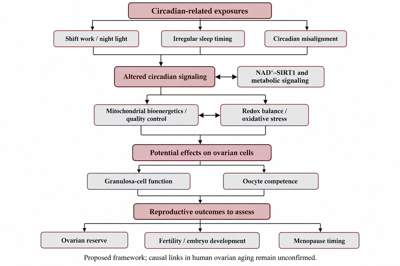
Figure 1: Integrative framework linking circadian disruption, mitochondrial dysfunction, and female reproductive aging. The original conceptual flowchart summarizes potential interactions among circadian exposures, metabolic signaling, mitochondrial bioenergetics, and redox homeostasis, and their possible consequences for ovarian-cell function and reproductive outcomes. Causal relationships in human ovarian aging remain to be established
PHYSIOLOGICAL FOUNDATIONS OF FEMALE REPRODUCTIVE AGING
Ovarian Reserve, Follicular Depletion, and Reproductive Lifespan: Aging of the female reproductive system is essentially related to the progressive depletion of the ovarian follicular pool. The human ovary is unique in comparison to most somatic tissues in that it has a finite number of primordial follicles, most of which were set aside prior to birth 10,11. Ovarian ageing is not just a decrease in the number of follicles. It also includes modifications in the developmental competence of surviving oocytes and in the physiological conditions under which follicular maturation takes place. Meiotic chromosome segregation abnormalities, mitochondrial activity, cellular stress responses, and follicular support mechanisms are all affected by age, which can affect the probability that an ovulated oocyte will give rise to a viable embryo 10,12. Thus, there are two aspects of reproductive ageing: loss of ovarian quantity and loss of oocyte quality. The clinical implications of the postmenopausal transition may extend beyond reproductive function to include cardiometabolic health. Observational research examining body mass index and lipid profiles in postmenopausal women provides a related clinical perspective on health assessment during this stage of life 72.
Oocyte Competence, Granulosa-Cell Function, and Steroidogenesis: The coordinated interaction of the oocyte with the somatic cells (granulosa and cumulus cells) is essential for successful follicular development. These cells contribute to the exchange of nutrients, metabolic support, signaling of growth factors, and the hormonal environment necessary for oocyte maturation. Granulosa cells are also involved in ovarian steroidogenesis, which involves their response to gonadotropins and involvement in the synthesis of both oestradiol and other reproductive hormones. The supportive activities may be affected by age-related changes in granulosa-cell metabolism and mitochondrial function 13, 14.The significance of these processes, however, varies from experimental model to experimental model and with the developmental stage of the follicles.
Chronological Age Versus Biological Ovarian Age: Chronological age is a useful indicator of reproductive age, but does not reflect the physiological diversity of ovarian function. Follicular reserves, reproductive endocrine profiles, and oocyte developmental competence may vary among women of the same age. These characteristics are also affected by genetic variation, environmental exposures, previous gonadotoxic treatment, and underlying reproductive disorders 12, 15. Ovarian function is also described by clinical measures, such as anti-Müllerian hormone (AMH), antral follicle count (AFC), and follicle-stimulating hormone (FSH), which are complementary. However, the measurement of ovarian reserve itself is mainly a measure of the number of follicles present and their response to ovarian stimulation, but not a measure of the quality of the oocytes or a reliable indicator of a woman's natural reproductive life 15. Therefore, biological ovarian age should not be viewed as a one-dimensional clinical parameter that has been validated. It is important to note this difference for the current review. These changes in ovarian physiology may occur through circadian disruption, mitochondrial dysfunction, and/or oxidative stress, but not necessarily at the same rates as other markers of ovarian ageing. Association with menstrual irregularity, such as an earlier age at menopause, does not prove accelerated follicular depletion.
Endocrine Aging Across the Reproductive Transition: Reproductive aging is also characterized by changes in hypothalamic, pituitary, and ovarian interactions. As the follicular reserve declines, ovarian feedback to the hypothalamus and pituitary changes, contributing to alterations in follicle-stimulating hormone and other reproductive hormones. These changes occur across the menopausal transition, which is characterized by increasing menstrual irregularity and ultimately the cessation of menstruation 16. Also relevant to this is the hypothalamic–pituitary–ovarian axis, as reproductive hormone secretion is also temporally regulated. Alterations in the level of hormones and their feedback regulation can affect ovarian function, and circadian patterns can regulate the timing of neuroendocrine activity. This gives a physiological framework for exploring the relationship between circadian misalignment and the process of reproductive aging, as well as the assumption that the ovary is independent of upstream timekeeping mechanisms.
One more potential connection between endocrine regulation and age-related changes in the ovary could be through mitochondrial dysfunction and oxidative damage. Recent studies have shown age-related changes in the energy metabolism, mitochondrial integrity, and redox balance of the oocyte, and studies of reduced ovarian reserve have found associations with changes in mitochondrial markers 17, 19. The results of these studies suggest that there is a need for additional research into mitochondrial and metabolic mechanisms, which do not necessarily mean that circadian disruption is the triggering source. Ovarian aging encompasses the progressive decrease in the size of the ovary, reduced competence of oocytes, ovarian somatic-cell changes, and age-related endocrine regulation.
CIRCADIAN RHYTHMS, SLEEP TIMING, AND REPRODUCTIVE ENDOCRINE REGULATION
Central Circadian Control: The suprachiasmatic nucleus (SCN) regulates the circadian control of sleep–wake cycles, metabolism, and endocrine function in response to environmental cues. It acts via neural and hormonal pathways on the hypothalamic–pituitary–gonadal axis (HPG axis), a system responsible for the regulation of follicular development, steroidogenesis, and ovulation 20,21. Experimental evidence shows that circadian signaling is involved in timing the release of reproductive hormones, such as the preovulatory surge of luteinizing hormone (LH). The mechanism is different in species, and the effects of circadian disruption on human ovarian ageing have not yet been established 21, 22.
Sleep Timing and Reproductive Function: There are three exposures: sleep duration, chronotype, and circadian alignment. Short sleep is an indication of lack of sleep opportunity, and social jet lag and irregular sleep schedules can result in a misalignment of the internal biological clock and external demands. These factors can overlap but should not be considered synonymous. A systematic review found associations between sleep disturbances and female reproductive outcomes, but the associations differed between populations and study designs 20. The findings don't confirm accelerated ovarian ageing, but in a prospective preconception cohort, sleep difficulties were associated with decreased fecundability 22. To better understand this association, more longitudinal research that assesses ovarian reserve and objective sleep and circadian markers is necessary. Human evidence from non-reproductive populations also provides context for the relationship between sleep disturbance and melatonin physiology. A study of emergency doctors examined insomnia prevalence and its relationship with serum melatonin, offering indirect evidence relevant to sleep–melatonin regulation 73. However, this study did not assess women-specific reproductive outcomes and cannot establish a link between sleep disruption and ovarian aging.
Circadian Regulation of Reproductive Hormones: The HPG axis regulates the activity of gonadotropin-releasing hormone, follicle-stimulating hormone (FSH), LH, and ovarian steroid hormones. Circadian signals can affect the hormone secretion rhythm and sensitivity of reproductive tissues 21. Disruption in menstrual regularity or ovulatory coordination may therefore occur without a decrease in the follicular reserve. Night-shift workers have been reported to have irregular periods, although this may be due to residual confounding by stress, metabolic health and sleep quality, and thus does not provide causal conclusions 23, 24. It is thus not a harbinger of ovarian ageing, but rather a possible reproductive stressor, associated with night-shift exposure.
Melatonin and Ovarian Hormone Signaling: Melatonin is a hormone that is involved in the perception of biological night and has been studied for its role in ovarian function and antioxidant activity 25. Experimental data indicate that it can affect follicular development and oocyte maturation, but this is dependent on physiologic context. The presence of antioxidant activity does not support the use of melatonin supplements to protect human ovarian reserve. Other studies have found that the reproductive hormones also affect sleep and thermoregulation, suggesting a possible bidirectional interaction between reproductive hormones and circadian physiology 26. These relationships make causal interpretation difficult since sleep changes can occur before, during, or after reproductive hormonal changes.
Shift Work and Reproductive Outcomes: Night-shift workers are exposed to a combination of several factors, such as nocturnal light, sleep disruption, and irregular eating patterns. There has been evidence of associations with reproductive outcomes in a few reviews, although exposure terms and results are not uniform in these studies 27. Requirements for future studies include differentiation of occupational schedules from objectively measured circadian misalignment and longitudinal study of ovarian reserve. The main evidence and its limitations are summarized in Table 1.
Table 1: Evidence linking circadian disruption and sleep-related exposures with female reproductive physiology
|
(Author et al., Year) Reference
|
Study design/population
|
Circadian exposure or mechanism
|
Main reproductive findings/relevance
|
Limitations
|
| Hu et al., 2023 4 Bracci et al., 20145 |
Meta-analysis |
Shift work |
Examines associations between shift work and menstrual irregularity |
Heterogeneity among included studies |
| Bracci et al., 20145 |
Observational study of rotating-shift nurses |
Peripheral clock-gene expression, melatonin, and estradiol |
Investigates changes in circadian and endocrine markers |
Limited generalizability |
| Stocker et al., 2014 6 |
Systematic review and meta-analysis |
Shift work |
Evaluates associations with early reproductive outcomes |
Limited mechanistic evidence |
| Caetano et al., 2021 20 |
Systematic review |
Sleep and reproductive function |
Reviews sleep-related reproductive outcomes |
Heterogeneous exposures and outcomes |
| Shao et al., 2021 21 |
Mechanistic review |
Circadian regulation of the hypothalamic–pituitary–gonadal (HPG) axis |
Describes central and peripheral clocks involved in reproductive regulation |
Several mechanisms derive from experimental models |
| Willis et al., 2019 22 |
Prospective preconception cohort |
Sleep patterns and shift work |
Sleep difficulties were associated with reduced fecundability; shift work was not associated in the same way |
Observational exposure assessment |
| Kim et al., 2024 23 |
Longitudinal cohort |
Night-shift work |
Investigates incident menstrual irregularity |
Does not directly measure ovarian aging |
| Lawson et al., 2011 24 |
Epidemiological study |
Rotating shift work |
Examines menstrual-cycle characteristics |
Potential residual confounding |
| Gamble et al., 2013 25 |
Mechanistic reviewy |
Circadian disruption |
Discusses possible effects on reproductive timing |
Does not establish direct causation |
Note: Fecundability refers to the probability of conception per menstrual cycle. Evidence of menstrual or fertility disturbance should not be interpreted as direct evidence of accelerated depletion of ovarian reserve
THE OVARIAN MOLECULAR CLOCK: CLOCK GENES AND FOLLICULAR PHYSIOLOGY
Core Circadian Clock Genes: A molecular circadian clock is regulated by a network of interconnected transcriptional feedback loops. CLOCK and BMAL1 are responsible for stimulating PER and CRY gene expression, which in turn inhibits CLOCK–BMAL1 activity. Other regulators, such as REV-ERB and ROR, help to control these oscillations and the downstream cellular processes 28, 29. Pathways that are clock-controlled may affect steroidogenesis, follicular development, and timing of ovulation in reproductive tissues. One measurement of clock-gene expression is not enough to confirm and quantify that an ovarian rhythm is disrupted; however, phase, amplitude, and temporal coordination should be taken into account.
Peripheral Clocks in Ovarian Tissues: Experimental research has shown that the expression of clock genes is found in ovarian cells, including granulosa cells. The expression of PER1 and BMAL1 shows follicular differences and gonadotropin responsiveness in rats, indicating ovarian clocks that are responsive to reproductive signals 31. Human evidence is preliminary. Brzezinski and coworkers demonstrated age-related changes in the expression of clock genes in in vitro fertilised luteinized granulosa cells 34.The results indicate a possible connection between ovarian ageing and clock regulation, but do not prove causality.
Clock Genes and Follicular Function: The activities of granulosa cells, including proliferation, hormone synthesis, and responsiveness to gonadotropins, may be controlled by clock genes. Estradiol production and the expression of the corresponding genes in bovine granulosa cells were modified by experimental suppression of Clock 30. Likewise, there were altered genes associated with steroidogenesis and prostaglandin synthesis in the reduced BMAL1-expressing rat luteinizing granulosa cells 32. Experimental evidence of local clock function in reproductive physiology was obtained by BMAL1 selective deletion of theca cells in mice, which disrupted ovulation 33.
Circadian Disruption and Ovarian Gene Expression: Circadian misalignment can affect the activity of the ovarian clock via modifications in the system hormones, neural, and metabolic cues. Environmental disruption is not a genetic elimination of an essential clock gene, however. If either exposure is to be assessed, it must be done through proper temporal measures and by using reproductive outcomes. A study of human luteinized granulosa cells reported an association between BMAL1 expression and genes involved in steroidogenesis, including STAR, CYP11A1, and CYP19A1 36.These results warrant further research but don't prove that sleep timing changes directly speed up the aging of the ovaries.
Bidirectional Interactions with Reproductive Hormones: There could be interactions between circadian regulation and reproductive hormones. Gonadotropin stimulation can alter the expression of the clock genes in the ovary, and pathways related to clock genes can influence the steroidogenic activity 29,31,35. Hormonal changes associated with age can thus affect the functioning of the ovarian clock, and conversely, changes in clock regulation could affect reproductive physiology.
MITOCHONDRIAL BIOENERGETICS AND THE AGING OVARY
Mitochondrial Energy Production and Oocyte Competence: Mitochondrial function supports oocyte maturation, fertilization, and early embryonic development through ATP production, calcium regulation, and metabolic signaling. Mitochondria produce ATP and control calcium signaling, metabolism, and production of reactive oxygen species (ROS). Therefore, the oocyte may be less competent in some aspects due to mitochondria-related alterations that occur with age and act in multiple interacting ways 38,39. Age-associated mitochondrial DNA damage and the limited capacity for its repair may further compromise mitochondrial function and oocyte competence 43. Any changes in mtDNA copy number should be assessed in the context of mitochondrial function, as an increased number of copies of mtDNA may only represent compensation for decreased bioenergetics and not increased cell health. Age-related mitochondrial dysfunction may compromise oocyte competence through impaired energy production, altered redox balance, and disrupted mitochondrial quality control Figure 2.
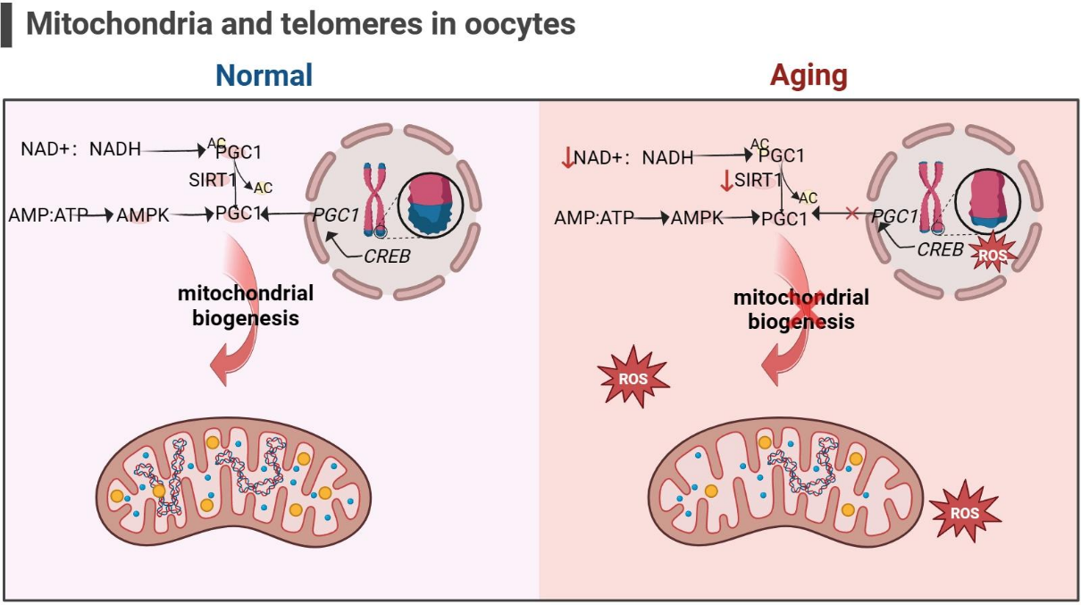
Figure 2: Mitochondrial dysfunction and telomere-associated mechanisms in oocyte aging 17
Membrane Potential, Calcium Regulation, and Meiotic Competence: The mitochondrial membrane potential is important for ATP production and for transport of metabolites. Disruption of this could impact cellular energy availability, calcium homeostasis, and may have an impact on meiotic progression and spindle organization. These processes are especially critical in oocytes, and abnormal segregation of the chromosomes is a significant factor in age-associated reproductive decline 37-40. Mitochondrial dysfunction is not the only reason for the occurrence of meiotic errors. Decreased female fertility in old age is also due to age-related alterations in the cohesins and other proteins involved in the regulation of the spindle apparatus, in addition to alterations in the quality of the oocytes 40.
Granulosa-Cell Mitochondria and Steroidogenesis: Mitochondrial function in granulosa and cumulus cells supports steroidogenesis and the metabolic environment required for follicular development 41. Age-related mitochondrial changes in these somatic cells may therefore influence oocyte competence indirectly, although the extent and timing of such effects in human reproductive aging remain incompletely defined.
Mitochondrial Quality Control and Mitophagy: Mitochondrial quality control is the process of removing damaged mitochondria and repairing and maintaining mitochondrial homeostasis. Removal of mitochondria by mitophagy selectively targets dysfunctional mitochondria, and the mitochondrial unfolded protein response is a mechanism for cells to respond to mitochondrial stress. There are indications that the quality-control mechanisms in the mitochondria may not function well in older oocytes. A recent study in 2023 indicated that the oocytes from aged females showed impaired mitochondrial stress-response and mitophagy mechanisms, which may be linked to decreased oocyte quality 42. These results provide a basis for the exploration of mitochondrial maintenance as a mechanism of reproductive ageing but do not yet prove to lead to a clinically validated technique for the restoration of human oocyte competence. In summary, mitochondrial dysfunction can influence the aging of reproduction by impacting energy metabolism, redox signaling, and organelle quality control Table 2 summarizes the principal mechanisms and limitations of this evidence.
Table 2: Mitochondrial Dysfunction and Cellular Mechanisms of Ovarian Aging
|
(Author et al., Year) Reference
|
Study design/model
|
Mitochondrial mechanism
|
Main findings/relevance to ovarian aging
|
Limitations
|
| Smits et al., 2023 7 |
Human ovarian tissue study |
Mitochondrial dysfunction and oxidative damage |
Reports age-associated oxidative damage and mitochondrial changes in human ovarian tissue |
Tissue findings do not establish causality |
| Alberico and Woods, 2022 13 |
Review |
Granulosa-cell metabolism |
Discusses mitochondrial and metabolic support of the follicular environment |
Evidence includes experimental models |
| Bao et al., 2024 14 |
Review |
Oocyte energy metabolism |
Reviews metabolic requirements and their relevance to ovarian aging |
Does not independently establish causal pathways |
| Ju et al., 2024 17 |
Review |
Mitochondrial dysfunction and potential interventions |
Summarizes mechanisms and candidate approaches relevant to ovarian aging |
Clinical effectiveness remains uncertain |
| van der Reest et al., 2021 37 |
Review |
Oocyte mitochondrial function |
Integrates mitochondrial energy production, redox regulation, and oocyte aging |
Evidence is drawn from heterogeneous models |
| Chiang et al., 2020 38 |
Review |
Mitochondrial dysfunction and reproductive longevity |
Discusses mitochondrial DNA, metabolism, and ovarian aging |
Relative contributions of mechanisms remain uncertain |
| Kasapoğlu and Seli, 2020 39 |
Review |
Mitochondrial dysfunction |
Examines potential links with ovarian aging and oocyte quality |
Therapeutic applications remain uncertain |
| Bebbere et al., 2022 40 |
Review |
Oocyte aging |
Connects cellular abnormalities with declining developmental competence |
Not focused exclusively on mitochondria |
| Khan et al., 2023 42 |
Experimental study |
Mitochondrial integrity and quality control |
Investigates mitochondrial integrity in relation to oocyte quality during reproductive aging |
Experimental findings require broader validation |
| Kobayashi and Imanaka, 2024 43 |
Review |
Mitochondrial DNA damage and repair in aging oocytes |
Examines how accumulated mitochondrial DNA damage and repair limitations may contribute to impaired energy production and oocyte dysfunction |
Mechanistic pathways and therapeutic implications require further validation |
|
Jiang and Shen, 2022 44
|
Review |
Mitochondrial therapeutic strategies |
Evaluates potential approaches for oocyte rescue |
Clinical efficacy remains uncertain |
|
Esencan et al., 2022 61
|
Narrative review |
Folliculogenesis and potential modifiers |
Discusses age-related changes in folliculogenesis and factors potentially affecting fertility outcomes |
Broad review; not specific to mitochondrial dysfunction |
|
Telfer et al., 2023 62
|
Review |
Oocyte health, aging, and development |
Discusses human oocyte quality and developmental capacity with age |
Covers several mechanisms beyond mitochondria |
Note: Mechanistic evidence from experimental systems should be distinguished from direct evidence in aging human ovaries. Association with oocyte aging does not by itself establish that mitochondrial dysfunction initiates reproductive decline.
CIRCADIAN–MITOCHONDRIAL CROSSTALK: AN INTEGRATED PHYSIOLOGICAL MECHANISM
Clock-Controlled Mitochondrial Metabolism: Every cell uses a clock to regulate metabolic shifts over the course of the day, enabling cells to prepare for shifts in energy needs. Metabolic pathways can be controlled by CLOCK–BMAL1 signaling and metabolites can modulate clock function. A key link is the mitochondria-specific nicotinamide adenine dinucleotide (NAD⁺) that is involved in mitochondrial metabolism and is required as a cofactor for the sirtuin enzymes 45,46. NAD⁺–SIRT1 pathway is a connection between metabolic status and clock control. CLOCK–BMAL1 affects the activity of NAMPT, an enzyme that affects the process of biosynthesis of NAD⁺, and NAD⁺-dependent SIRT1 changes the activity of proteins associated with the clock and metabolic regulators 45,47.
NAD⁺–SIRT1, AMPK, and PGC-1α Signaling: AMP-activated protein kinase (AMPK) is activated by a decrease in cellular energy, and peroxisome proliferator-activated receptor gamma coactivator 1-alpha (PGC-1α) regulates mitochondrial biogenesis and oxidative metabolism. SIRT1, AMPK and PGC-1α are linked in intricate metabolic pathways that regulate energy balance and mitochondrial function 46,48. These pathways could be important in reproductive ageing as the synchronised energy metabolism of oocytes and granulosa cells is essential. However, it should not be inferred from their known involvement in general cellular metabolism that circadian disruption significantly affects these pathways to accelerate human ovarian ageing.
Melatonin, Mitochondrial Function, and Redox Balance: Melatonin has been investigated for its effects on circadian signaling, antioxidant defences, and mitochondrial function in ovarian models 49,50. A 2025 study in a polycystic ovary syndrome (PCOS) model examined melatonin-related modulation of ovarian mitochondrial dysfunction involving the clock gene CLOCK 51. These findings provide mechanistic evidence in a pathological model, but they do not establish that melatonin prevents physiological ovarian aging or extends reproductive lifespan in humans.
Temporal Misalignment and Mitochondrial Stress: Circadian misalignment can disrupt the timing of metabolic need, substrate utilization, and cell repairs. From general circadian studies, it is clear that the metabolism of NAD(P)(H) and redox levels are coupled to clock activity and might explain how disruptions to the timing could be linked to mitochondrial homeostasis 47, 48. Such effects might potentially impact ATP availability, ROS handling, and mitochondrial quality control in ovarian cells. But there is so far little direct evidence linking sleep timing to these changes in aging human ovaries. Therefore, the proposed relationship should be considered a physiological hypothesis rather than a causal sequence.
Reciprocal Signaling Between Mitochondrial Dysfunction and the Circadian Clock: The metabolic pathways can be regulated by circadian regulation, and the cellular energy status and redox balance can modify clock-associated signaling. A bidirectional interaction occurs, whereby compromised mitochondrial functions may impact cell timing, and disrupted cell timing may impact mitochondrial function 45,48. The resulting model is not a linear progression from poor sleep to damage to the mitochondria to ovarian ageing, but rather a model of interacting processes. The proposed interaction between circadian regulation, mitochondrial metabolism, and redox homeostasis provides a framework for investigating their potential contribution to ovarian aging, although direct evidence in human ovaries remains limited 45-50. The potential involvement of ovarian clock signaling in mitochondrial dysfunction is also supported by experimental findings from a PCOS model 51. These interactions are illustrated in Figure 3.
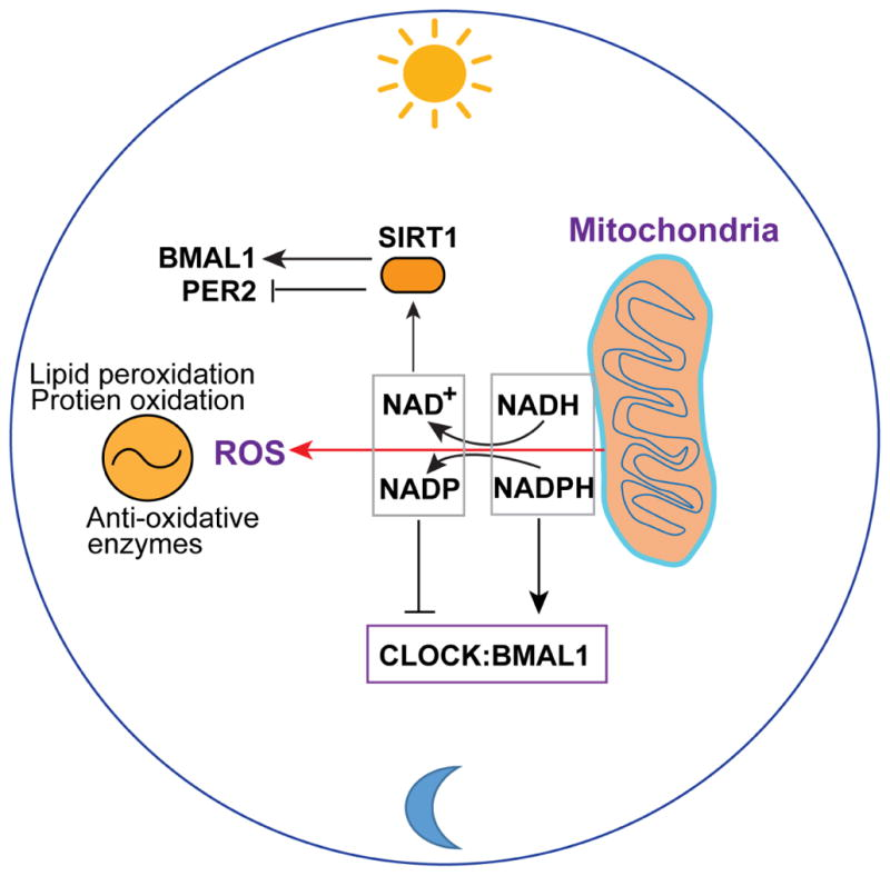
Figure 3: Circadian regulation, mitochondrial metabolism, and oxidative stress 71
Table 3 summarizes the relevant molecular links and their evidential limitations.
Table 3: Molecular Links Between Circadian Regulation, Mitochondrial Function, and Ovarian Aging
|
(Author et al., Year) Reference
|
Study design
|
Circadian regulator or pathway
|
Mitochondrial / redox mechanism
|
Relevance and evidence limitations
|
| Bellet et al., 2011 45 |
Mechanistic review |
NAD⁺–SIRT1 signaling and the circadian clock |
Links cellular metabolic state with clock-associated regulation |
General metabolic evidence; not ovary-specific |
| Eckel-Mahan and Sassone-Corsi, 201346 |
Mechanistic review |
Reciprocal circadian and metabolic regulation |
Describes interactions between clock activity and metabolic pathways |
Much of the evidence comes from non-reproductive systems |
| Levine et al., 2022 47 |
Review |
Circadian NAD(P)(H) cycles |
Describes temporal regulation of cellular redox metabolism |
Does not establish effects on ovarian aging |
| Asher and Schibler, 2011 48 |
Mechanistic review |
Crosstalk between circadian and metabolic cycles |
Explains reciprocal regulation of metabolic and clock-associated processes |
General mammalian evidence; direct ovarian effects remain uncertain |
| Jiang et al., 2021 49 |
Review |
Melatonin and reproductive oxidative stress |
Reviews melatonin-associated antioxidant and reproductive mechanisms |
Does not establish prevention of physiological ovarian aging |
| Reiter et al., 2023 50 |
Review |
Melatonin in age-related ovarian failure |
Discusses possible roles in oxidative balance and reproductive function |
Proposed benefits require stronger clinical evidence |
| Chen et al., 2025 51 |
Experimental study in a polycystic ovary syndrome (PCOS) model |
CLOCK-associated regulation and melatonin |
Examines melatonin-related changes in ovarian mitochondrial dysfunction |
PCOS is not equivalent to physiological ovarian aging |
Note: These studies support a plausible circadian–metabolic interaction, not a proven causal pathway from disrupted sleep to mitochondrial damage and accelerated human ovarian aging. Findings from PCOS models should not be generalized directly to physiological reproductive aging.
OXIDATIVE STRESS, REDOX IMBALANCE, AND FOLLICULAR DECLINE
Reactive Oxygen Species and Physiological Redox Signaling: Reactive oxygen species (ROS) are involved in cellular signaling, follicular development, and the ovulatory process. If the production of ROS is greater than the capacity of antioxidants and repair mechanisms, then oxidative stress may cause damage to cell components and interfere with ovarian function 53-55. Mitochondrial dysfunction with age could also add to the oxidative load and further affect oocyte competence and follicular degeneration.
Oxidative Damage to Mitochondrial Components: Excessive ROS can damage mitochondrial DNA, oxidize proteins, and disrupt membrane lipids, potentially impairing respiratory-chain activity and ATP production 54-56. As people get older, they may be more vulnerable to oxidative damage due to these changes in defences. However, ROS also play a critical role in normal physiological processes, and indiscriminate suppression may disrupt normal reproductive signaling.
Mitochondrial Damage and Follicular Loss: Oxidative stress may damage mitochondria, activate cellular stress responses, and promote apoptosis, potentially contributing to granulosa-cell dysfunction and follicular atresia 53,59. There may also be interactions between oxidative damage and inflammation, and DNA damage and cellular senescence, which are more prominent for different types of ovarian cells at different developmental stages.
Feedback Between Oxidative Stress and Circadian Disruption: There are links between circadian clocks and aspects of metabolism and antioxidant defense, and cellular redox state can affect clock-related signaling 56,60. These interactions could be used as a plausible mechanism to explain how circadian misalignment and mitochondrial dysfunction might feed into each other. This feedback loop is demonstrated directly in aging human ovaries with only limited evidence. Oxidative stress is therefore considered a possible mechanism linking mitochondrial dysfunction to follicular decline, but not as a direct result of sleep timing disruption.
CONSEQUENCES FOR OVARIAN RESERVE, FERTILITY, AND REPRODUCTIVE LONGEVITY
Follicular Reserve and Reproductive Aging: Ovarian reserve is indicative of the number of follicles left, and reproductive aging is a complex process involving both the number and quality of follicles. There is a similarity between these dimensions, but they are not synonymous 10,11. Several changes in the ovarian environment, mitochondrial function, and cell maintenance, all of which likely play a role in reproductive aging, may be affected by increasing age, but their individual contribution is not completely elucidated 61.
Oocyte Quality and Embryo Development: Maternal age is linked to increased meiotic errors and chromosomal aneuploidy, which leads to loss of embryo developmental competence, infertility, and miscarriage 62,63. Other factors such as mitochondrial dysfunction, oxidative damage, and alteration of the follicular microenvironment may also impede further maturation of the oocyte. No single pathway is responsible for reproductive decline, which occurs through these mechanisms in addition to age-related changes in chromosome cohesion and spindle organization.
Endocrine Function and the Menopausal Transition: As the follicular pool declines, ovarian endocrine activity changes, contributing to menstrual irregularity and eventually menopause. This transition is a gradual process, which can not be reliably estimated by a single measurement of ovarian reserve. The physiological process of reproductive aging needs to be differentiated from the ovarian reserve markers in clinical assessment 15,16.
Biological Age and Reproductive Resilience: Genetic, metabolic, environmental, and cellular maintenance factors may contribute to interindividual variation in reproductive aging 62,64. Circadian disruption, mitochondrial dysfunction, and oxidative stress are plausible contributors, but their independent effects on human reproductive lifespan remain uncertain. Ovarian reserve, oocyte competence, fertility outcomes, and age at menopause should be evaluated separately because they represent related but distinct dimensions of reproductive function.
TRANSLATIONAL IMPLICATIONS AND FUTURE RESEARCH
Clinical Interpretation and Reproductive Counselling: Circadian disruption, mitochondrial dysfunction, and oxidative stress are biologically plausible contributors to variation in reproductive function, but their clinical significance for ovarian aging remains uncertain. Menstrual irregularity, sleep disturbance, and reduced fecundability should not be interpreted as direct evidence of accelerated follicular depletion. Anti-Müllerian hormone (AMH), antral follicle count (AFC), and follicle-stimulating hormone (FSH) provide complementary information about ovarian reserve but do not directly measure mitochondrial function or reliably determine an individual's natural reproductive lifespan 15,65.
Priorities for Mechanistic and Longitudinal Research: Future studies should examine the differences among sleep duration, chronotype, light exposure at night, and the objectively measured circadian misalignment. These measures should be used in prospective human cohorts in addition to repeated measurement of menstrual characteristics, reproductive hormones, AMH, AFC, and fertility outcomes. Where feasible, follicular-fluid metabolomics and mitochondrial-function markers could help identify changes in the ovarian microenvironment associated with circadian disruption. These methods would be based on the evidence of age-related oxidative damage in human ovarian tissue and mitochondrial metabolism alterations and would overcome the drawbacks of cross-sectional studies and ovarian-stimulation samples 7,66,69.
Additional molecular signatures of reproductive ageing may be discovered through multi-omics analysis, but candidate biomarkers will need to be validated separately in populations and age groups 67. Experimental models should be used to determine if alterations in clock-gene activity precede mitochondrial dysfunction or if the alterations in cellular metabolism are the result of mitochondrial dysfunction. The distinction of physiological reproductive aging from pathological conditions such as premature ovarian insufficiency and polycystic ovary syndrome is also important since mechanisms seen in these states may not apply to healthy ovarian aging, and should be considered 68.
Priorities for Mechanistic and Longitudinal Research: Regularity of sleep, proper light exposure and healthier lifestyle patterns are good general health measures but evidence to support the maintenance of ovarian reserve or extension of reproductive lifespan is lacking. Melatonin, antioxidant supplements, mitochondrial-targeted compounds, and metabolic modulators have been investigated as potential interventions in selected experimental or clinical contexts. Although improvements in oxidative balance or the quality of the oocytes have been observed in preclinical trials, their clinical benefits, long-term safety, and improved live-birth rates remain unknown 69,70. Future studies using interventions are recommended to employ well-defined reproductive outcomes, control groups, an appropriate follow-up time period, and explicit evaluation of adverse effects. The first major step is to determine whether there is a measurable impact of modifiable circadian exposures on ovarian biology, and to develop targeted interventions based on this.
CONCLUSION
Female reproductive aging involves progressive follicular depletion, declining oocyte developmental competence, and changes in reproductive endocrine function. Circadian regulation, mitochondrial bioenergetics, and oxidative stress may interact through pathways governing cellular energy balance and redox homeostasis, offering a plausible framework for investigating variation in reproductive aging beyond chronological age. However, direct evidence that circadian disruption accelerates ovarian reserve depletion or shortens reproductive lifespan in humans remains limited. Longitudinal studies integrating objective circadian measures, ovarian reserve markers, reproductive outcomes, and mitochondrial assessments are needed to establish the direction and clinical significance of these relationships. At present, the circadian–mitochondrial framework should be considered a testable physiological hypothesis rather than an established causal pathway or a basis for claiming that specific interventions preserve reproductive longevity.
References
- Dong L, Teh DB, Kennedy BK, Huang Z. Unraveling female reproductive senescence to enhance healthy longevity. Cell Res. 2023;33(1):11-29. https://doi.org/10.1038/s41422-022-00718-7
- Das A, Destouni A. Novel insights into reproductive ageing and menopause from genomics. Hum Reprod. 2023;38(2):195-203. https://doi.org/10.1093/humrep/deac256
- Yaw AM, McLane-Svoboda AK, Hoffmann HM. Shiftwork and light at night negatively impact molecular and endocrine timekeeping in the female reproductive axis in humans and rodents. Int J Mol Sci. 2020;22(1):324. https://doi.org/10.3390/ijms22010324
- Hu F, Wu C, Jia Y, Zhen H, Cheng H, Zhang F, et al. Shift work and menstruation: A meta-analysis study. SSM Popul Health. 2023;24:101542. https://doi.org/10.1016/j.ssmph.2023.101542
- Bracci M, Manzella N, Copertaro A, Staffolani S, Strafella E, Barbaresi M, et al. Rotating-shift nurses after a day off: peripheral clock gene expression, urinary melatonin, and serum 17-β-estradiol levels. Scand J Work Environ Health. 2014:295-304. https://doi.org/10.5271/sjweh.3414
- Stocker LJ, Macklon NS, Cheong YC, Bewley SJ. Influence of Shift Work on Early Reproductive Outcomes: A Systematic Review and Meta-analysis. Obstet Gynecol. 2014;124(1):99-110. https://doi.org/10.1097/AOG.0000000000000321
- Smits MA, Schomakers BV, van Weeghel M, Wever EJ, Wüst RC, Dijk F, et al. Human ovarian aging is characterized by oxidative damage and mitochondrial dysfunction. Hum Reprod. 2023;38(11):2208-20. https://doi.org/10.1093/humrep/dead177
- Liu Y, Gao J. Reproductive aging: biological pathways and potential interventive strategies. J Genet Genomics. 2023;50(3):141-50. https://doi.org/10.1016/j.jgg.2022.07.002
- Wang X, Wang L, Xiang W. Mechanisms of ovarian aging in women: a review. J Ovarian Res. 2023;16(1):67. https://doi.org/10.1186/s13048-023-01151-z
- Broekmans FJ, Soules MR, Fauser BC. Ovarian aging: mechanisms and clinical consequences. Endocr Rev. 2009;30(5):465-93. https://doi.org/10.1210/er.2009-0006
- Wallace WH, Kelsey TW. Human ovarian reserve from conception to the menopause. PLoS One. 2010;5(1):e8772. https://doi.org/10.1371/journal.pone.0008772
- Te Velde ER, Pearson PL. The variability of female reproductive ageing. Hum Reprod Update. 2002;8(2):141-54. https://doi.org/10.1093/humupd/8.2.141
- Alberico HC, Woods DC. Role of granulosa cells in the aging ovarian landscape: a focus on mitochondrial and metabolic function. Front Physiol. 2022;12:800739. https://doi.org/10.3389/fphys.2021.800739
- Bao S, Yin T, Liu S. Ovarian aging: energy metabolism of oocytes. J Ovarian Res. 2024;17(1):118. https://doi.org/10.1186/s13048-024-01427-y
- Practice Committee of the American Society for Reproductive Medicine. Testing and interpreting measures of ovarian reserve: a committee opinion. Fertil Steril. 2015;103(3):e9-17. https://doi.org/10.1016/j.fertnstert.2020.09.134
- Harlow SD, Gass M, Hall JE, Lobo R, Maki P, Rebar RW, et al. Executive summary of the Stages of Reproductive Aging Workshop+ 10: addressing the unfinished agenda of staging reproductive aging. Climacteric. 2012;15(2):105-14. https://doi.org/10.1097/gme.0b013e31824d8f40
- Ju W, Zhao Y, Yu Y, Zhao S, Xiang S, Lian F. Mechanisms of mitochondrial dysfunction in ovarian aging and potential interventions. Front Endocrinol. 2024;15:1361289. https://doi.org/10.3389/fendo.2024.1361289
- Yang Z, Liu S, Pan X. Research progress on mitochondrial damage and repairing in oocytes: A review. Mitochondrion. 2024;75:101845. https://doi.org/10.1016/j.mito.2024.101845
- Zhu Q, Li Y, Ma J, Ma H, Liang X. Potential factors result in diminished ovarian reserve: a comprehensive review. J Ovarian Res. 2023;16(1):208. https://doi.org/10.1186/s13048-023-01296-x
- Caetano G, Bozinovic I, Dupont C, Léger D, Lévy R, Sermondade N. Impact of sleep on female and male reproductive functions: a systematic review. Fertil Steril. 2021;115(3):715-31. https://doi.org/10.1016/j.fertnstert.2020.08.1429
- Shao S, Zhao H, Lu Z, Lei X, Zhang Y. Circadian rhythms within the female HPG axis: from physiology to etiology. Endocrinology. 2021;162(8):bqab117. https://doi.org/10.1210/endocr/bqab117
- Willis SK, Hatch EE, Wesselink AK, Rothman KJ, Mikkelsen EM, Wise LA. Female sleep patterns, shift work, and fecundability in a North American preconception cohort study. Fertil Steril. 2019;111(6):1201-10. https://doi.org/10.1016/j.fertnstert.2019.01.037
- Kim K, Lee MY, Chang Y, Ryu S. Nightshift work and irregular menstrual cycle: 8-year follow-up cohort study. Occup Med. 2024;74(2):152-60. https://doi.org/10.1093/occmed/kqad162
- Lawson CC, Whelan EA, Hibert EN, Spiegelman D, Schernhammer ES, Rich-Edwards JW. Rotating shift work and menstrual cycle characteristics. Epidemiology (Cambridge, Mass.). 2011;22(3):305. https://doi.org/10.1097/EDE.0b013e3182130016
- Gamble KL, Resuehr D, Johnson CH. Shift work and circadian dysregulation of reproduction. Front Endocrinol (Lausanne). 2013;4:92. https://doi.org/10.3389/fendo.2013.00092
- Alzueta E, Baker FC. The menstrual cycle and sleep. Sleep Med Clin. 2023;18(4):399-413. https://doi.org/10.1016/j.jsmc.2023.06.003
- Fernandez RC, Marino JL, Varcoe TJ, Davis S, Moran LJ, Rumbold AR, et al. Fixed or rotating night shift work undertaken by women: implications for fertility and miscarriage. Semin Reprod Med. 2016;34(2):74-82. https://doi.org/10.1055/s-0036-1571354
- Sellix MT, Menaker M. Circadian clocks in the ovary. Trends Endocrinol Metab. 2010;21(10):628-36. https://doi.org/10.1016/j.tem.2010.06.002
- Karman BN, Tischkau SA. Circadian clock gene expression in the ovary: effects of luteinizing hormone. Biol Reprod. 2006;75(4):624-32. https://doi.org/10.1095/biolreprod.106.050732
- Shimizu T, Hirai Y, Murayama C, Miyamoto A, Miyazaki H, Miyazaki K. Circadian Clock genes Per2 and clock regulate steroid production, cell proliferation, and luteinizing hormone receptor transcription in ovarian granulosa cells. Biochem Biophys Res Commun. 2011;412(1):132-5. https://doi.org/10.1016/j.bbrc.2011.07.058
- Gräs S, Georg B, Jørgensen HL, Fahrenkrug J. Expression of the clock genes Per1 and Bmal1 during follicle development in the rat ovary. Effects of gonadotropin stimulation and hypophysectomy. Cell Tissue Res. 2012;350(3):539-48. https://doi.org/10.1007/s00441-012-1489-2
- Chen H, Zhao L, Kumazawa M, Yamauchi N, Shigeyoshi Y, Hashimoto S, et al. Downregulation of core clock gene Bmal1 attenuates expression of progesterone and prostaglandin biosynthesis-related genes in rat luteinizing granulosa cells. Am J Physiol Cell Physiol. 2013;304(12):C1131-40. https://doi.org/10.1152/ajpcell.00008.2013
- Mereness AL, Murphy ZC, Forrestel AC, Butler S, Ko C, Richards JS, et al. Conditional deletion of Bmal1 in ovarian theca cells disrupts ovulation in female mice. Endocrinology. 2016;157(2):913-27. https://doi.org/10.1210/en.2015-1645
- Brzezinski A, Saada A, Miller H, Brzezinski-Sinai NA, Ben-Meir A. Is the aging human ovary still ticking?: Expression of clock-genes in luteinized granulosa cells of young and older women. J Ovarian Res. 2018;11(1):95. https://doi.org/10.1186/s13048-018-0471-3
- Urlep Z, Rozman D. The interplay between circadian system, cholesterol synthesis, and steroidogenesis affects various aspects of female reproduction. Front Endocrinol. 2013;4:111. https://doi.org/10.3389/fendo.2013.00111
- Kawamura T, Dai Y, Ono M, Kikuchi T, Yamanaka A, Ueno K, et al. BMAL1 positively correlates with genes regulating steroidogenesis in human luteinized granulosa cells. Reproduction. 2024;167(2):e230225. https://doi.org/10.1530/REP-23-0225
- van der Reest J, Cecchino GN, Haigis MC, Kordowitzki P. Mitochondria: Their relevance during oocyte ageing. Ageing Res Rev. 2021;70:101378. https://doi.org/10.1016/j.arr.2021.101378
- Chiang JL, Shukla P, Pagidas K, Ahmed NS, Karri S, Gunn DD, et al. Mitochondria in ovarian aging and reproductive longevity. Ageing Res Rev. 2020;63:101168. https://doi.org/10.1016/j.arr.2020.101168
- Kasapoğlu I, Seli E. Mitochondrial dysfunction and ovarian aging. Endocrinology. 2020;161(2):bqaa001. https://doi.org/10.1210/endocr/bqaa001
- Bebbere D, Coticchio G, Borini A, Ledda S. Oocyte aging: looking beyond chromosome segregation errors. J Assist Reprod Genet. 2022;39(4):793-800. https://doi.org/10.1007/s10815-022-02441-z
- Sreerangaraja Urs DB, Wu WH, Komrskova K, Postlerova P, Lin YF, Tzeng CR, et al. Mitochondrial function in modulating human granulosa cell steroidogenesis and female fertility. Int J Mol Sci. 2020;21(10):3592. https://doi.org/10.3390/ijms21103592
- Khan SA, Reed L, Schoolcraft WB, Yuan Y, Krisher RL. Control of mitochondrial integrity influences oocyte quality during reproductive aging. Mol Hum Reprod. 2023;29(9):gaad028. https://doi.org/10.1093/molehr/gaad028
- Kobayashi H, Imanaka S. Mitochondrial DNA Damage and Its Repair Mechanisms in Aging Oocytes. Int J Mol Sci. 2024;25(23):13144. https://doi.org/10.3390/ijms252313144
- Jiang Z, Shen H. Mitochondria: emerging therapeutic strategies for oocyte rescue. Reprod Sci. 2022;29(3):711-22. https://doi.org/10.1007/s43032-021-00523-4
- Bellet MM, Orozco-Solis R, Sahar S, Eckel-Mahan K, Sassone-Corsi P. The time of metabolism: NAD+, SIRT1, and the circadian clock. Cold Spring Harb Symp Quant Biol. 2011;76:31-38. https://doi.org/10.1101/sqb.2011.76.010520
- Eckel-Mahan K, Sassone-Corsi P. Metabolism and the Circadian Clock Converge. Physiol Rev. 2013;93:107-135. https://doi.org/10.1152/physrev.00016.2012
- Levine DC, Ramsey KM, Bass J. Circadian NAD (P)(H) cycles in cell metabolism. Semin Cell Dev Biol. 2022;126:15-26. https://doi.org/10.1016/j.semcdb.2021.07.008
- Asher G, Schibler U. Crosstalk between components of circadian and metabolic cycles in mammals. Cell Metab. 2011;13(2):125-37. https://doi.org/10.1016/j.cmet.2011.01.006
- Jiang Y, Shi H, Liu Y, Zhao S, Zhao H. Applications of melatonin in female reproduction in the context of oxidative stress. Oxid Med Cell Longev. 2021;2021(1):6668365. https://doi.org/10.1155/2021/6668365
- Reiter RJ, Sharma R, Romero A, Manucha W, Tan DX, Zuccari DA, et al. Aging-related ovarian failure and infertility: melatonin to the rescue. Antioxidants. 2023;12(3):695. https://doi.org/10.3390/antiox12030695
- Chen W, Zhang H, Guo B, Tao Y, Zhang J, Wang J, et al. Melatonin refines ovarian mitochondrial dysfunction in PCOS by regulating the circadian rhythm gene Clock: W. Cell Mol Life Sci. 2025;82(1):104. https://doi.org/10.1007/s00018-025-05609-9
- Panda S. Circadian physiology of metabolism. Science. 2016;354(6315):1008-15. https://doi.org/10.1126/science.aah4967
- Yan F, Zhao Q, Li Y, Zheng Z, Kong X, Shu C, et al. The role of oxidative stress in ovarian aging: a review. J Ovarian Res. 2022;15(1):100. https://doi.org/10.1186/s13048-022-01032-x
- Wang L, Tang J, Wang L, Tan F, Song H, Zhou J, Li F. Oxidative stress in oocyte aging and female reproduction. J Cell Physiol. 2021;236(12):7966-83. https://doi.org/10.1002/jcp.30468
- Martin JH, Nixon B, Cafe SL, Aitken RJ, Bromfield EG, Lord T. Oxidative stress and reproductive function: oxidative stress and in vitro ageing of the post-ovulatory oocyte: an update on recent advances in the field. Reproduction. 2022;164(6):F109-24. https://doi.org/10.1530/REP-22-0206
- Di Nisio V, Antonouli S, Damdimopoulou P, Salumets A, Cecconi S, Sierr. In vivo and in vitro postovulatory aging: when time works against oocyte quality? J Assist Reprod Genet. 2022;39(4):905-918. https://doi.org/10.1007/s10815-022-02418-y
- Ansere VA, Ali-Mondal S, Sathiaseelan R, Garcia DN, Isola JV, Henseb JD, et al. Cellular hallmarks of aging emerge in the ovary prior to primordial follicle depletion. Mech Ageing Dev. 2021;194:111425. https://doi.org/10.1016/j.mad.2020.111425
- Tesarik J, Galán-Lázaro M, Mendoza-Tesarik R. Ovarian aging: molecular mechanisms and medical management. Int J Mol Sci. 2021;22(3):1371. https://doi.org/10.3390/ijms22031371
- Jadeja RN, Martin PM, Chen W. Mitochondrial oxidative stress and energy metabolism: impact on aging and longevity. Oxid Med Cell Longev. 2021;2021:9789086. https://doi.org/10.1155/2021/9789086
- Di Emidio G, Falone S, Artini PG, Amicarelli F, D’Alessandro AM, Tatone C. Mitochondrial sirtuins in reproduction. Antioxidants. 2021;10(7):1047. https://doi.org/10.3390/antiox10071047
- Esencan E, Beroukhim G, Seifer DB. Age-related changes in Folliculogenesis and potential modifiers to improve fertility outcomes-A narrative review. Reprod Biol Endocrinol. 2022;20(1):156. https://doi.org/10.1186/s12958-022-01033-x
- Telfer EE, Grosbois J, Odey YL, Rosario R, Anderson RA. Making a good egg: human oocyte health, aging, and in vitro development. Physiol Rev. 2023;103(4):2623-77. https://doi.org/10.1152/physrev.00032.2022
- Charalambous C, Webster A, Schuh M. Aneuploidy in mammalian oocytes and the impact of maternal ageing. Nat Rev Mol Cell Biol. 2023;24(1):27-44. https://doi.org/10.1038/s41580-022-00517-3
- Zhu Z, Xu W, Liu L. Ovarian aging: mechanisms and intervention strategies. Med Rev. 2023;2(6):590-610. https://doi.org/10.1515/mr-2022-0031
- Ono M, Dai Y, Fujiwara T, Fujiwara H, Daikoku T, Ando H, et al. Influence of lifestyle and the circadian clock on reproduction. Reprod Med Biol. 2025;24:e12641. https://doi.org/10.1002/rmb2.12641
- Ingole S, Khare K, Dwivedi V, Taksande B, Umekar M, Mangrulkar S. Decoding ovarian aging in women: Cellular damage, signaling networks, and treatment frontiers. Reprod Biol. 2025;25(4):101075. https://doi.org/10.1016/j.repbio.2025.101075
- Wang R, Li Y, Zhu L. Advances in multi-omics and aging clock research for female reproductive health and aging. MedScience. 2026;20:94-119. https://doi.org/10.1007/s11684-026-1207-1
- Fang Q, Qiao Z, Li F, Bai S, Chen M, Chen S, et al. Melatonin Alleviates Sleep Disturbance-Induced Ovarian Reserve Decline by Suppressing NLRP3-Mediated Pyroptosis in Granulosa Cells. J Pineal Res. 2026;78(4):e70151. https://doi.org/10.1111/jpi.70151
- Kobayashi H, Imanaka S. Exploring potential pathways from oxidative stress to ovarian aging. J Obstet Gynaecol Res. 2025;51(1):e16166. https://doi.org/10.1111/jog.16166
- Mondal R, Nandi M. Ovarian ageing: from biological constraint to emerging therapeutic frontier. J Assist Reprod Genet. 2026. https://doi.org/10.1007/s10815-026-04011-z
- Yang Z, Kim H, Ali A, Zheng Z, Zhang K. Interaction between stress responses and circadian metabolism in metabolic disease. Liver Res (Beijing, China). 2017;1(3):156-62. https://doi.org/10.1016/j.livres.2017.11.002
- Lohana K, Parkash J, Shaikh MZ, Ghani M, Lohana N, Uqaili AA. To deduce the changes in lipid profile and BMI that lead to cardiovascular diseases in postmenopausal women. J Popul Ther Clin Pharmacol. 2023;30(19):1809-1814. https://doi.org/10.53555/jptcp.v30i19.3941
- Dars JJ, Memon SF, Uqaili AA, Shaikh S, Tagar M, Javed S. Prevalence of insomnia among emergency doctors and their physiological link with serum melatonin: Key to better sleep among doctors of LUMHS. Pak J Med Health Sci. 2022;16(9):664. https://doi.org/10.53350/pjmhs22169664
This work is published by Multidisciplinary Scholarly Advancement and Research MSAR Institute. © 2026 The Author(s). The full terms of Journal Publishing policies are available at https://msarjournals.com/index.php/index/open-access and incorporate the Creative Commons Attribution – Non Commercial (CC BY-NC 4.0) License https://creativecommons.org/licenses/by-nc/4.0/. By accessing the work you hereby accept the Terms. Non-commercial uses of the work are permitted without any further permission, provided the work is properly attributed. Publisher’s Note: MSAR Institute remains neutral with regard to jurisdictional claims in published maps and institutional affiliations, and assumes no liability for the scientific accuracy or clinical efficacy of the content herein, as they rest entirely with the authors.